

A - negative (red) RAF, XYL and MDG tests when
incubated aerobically;
B - false-positive (yellow) RAF, XYL and MDG tests
when incubated in 20% CO2 atmosphere
incubated aerobically;
B - false-positive (yellow) RAF, XYL and MDG tests
when incubated in 20% CO2 atmosphere
| A - alpha-hemolysis, B - beta-hemolysis, C - false hemolysis produced by acidic inoculum |
| Catalase test: A - positive (Staphylococus aureus), B - false positive (no culture, traces of blood agar on loop) |
| Indole production test: A - positive, B - weak positive, C - false positive |
(c) Costin Stoica
| Antibiogram |
| Encyclopedia |
| Culture media |
| Biochemical tests |
| Stainings |
| Images |
| Movies |
| Articles |
| Identification |
| Software |
| R E G N U M PROKARYOTAE |

| Back |
| False Results of Biochemical Tests |
Example 1 - pigment production
Klebsiella oxytoca strains can produce a dark
brown pigment when growth on media containing
gluconate and ferric citrate. Klebsiella does not
produce hydrogen sulfide and hence does not
produce a dark ring when tested on TSI agar. In
this case the TSI slant is darkened by pigment,
not by hydrogen sulfide production.
Example 2 - pigment on chromogenic media
MacConkey Agar is frequently used as selective
media for the Enterobacteriaceae. The growth of
Gram-positive germs is inhibited by the bile salts
and the crystal violet. Medium also contains
lactose and neutral red as pH indicator.
Lactose-fermenting germs form red colonies,
lactose-negative germs form colorless colonies.
Serratia marcescens does not ferment lactose,
hence it should form colorless colonies. But this
species produce a red pigment resulting red
colonies on MacConkey agar which may be
taken as lactose-positive.
Example 3 - old culture
With very few exceptions, Salmonella is a
microorganism that does not ferment lactose.
Therefore, when grown on MacConkey, it
produces colorless colonies.
In the presented case, following a prolonged
storage in the refrigerator (more than a month),
the environment around the colonies became
acidic and caused the red color change of the
colonies.
Example 4 - blood interference
Catalase is an enzyme present in most of the
organisms and is involved in decomposition of
the hydrogen peroxyde in H2O and O2. If
catalase is present, bubbles will form from the
oxygen that is made in the reaction: 2H2O2 +
catalase => 2H2O + O2 + catalase. Test is
negative if effervescence is not produced.
But not only bacteria can produce catalase;
media containing whole red blood cells will
contain catalase and could therefore give a
false positive result. If using colonies from a
blood agar plate, be very careful not to scrape
up any of the blood agar.
Example 5 - slime
Like the Gram stain reaction, the potassium
hydroxide test is based on differences in the
bacterial wall composition. The cell wall of
Gram-negative bacteria is easily disrupted when
exposed to dilute alkali solutions while the tough
thick peptidoglycan wall of Gram-positives does
not lyse. When the cell walls are disrupted, the
suspension in KOH becomes viscous due to the
release of relatively unfragmented threads of
deoxyribonucleic acid. But, some Gram-positive
species like Bacillus subtilis can produce a
viscous substance / slime resembling the DNA
released from Gram-negatives in KOH solution.
Example 6 - bad neighbours
The results of reactions based on color change
following acidification or alkalinization of the
environment can be falsified by the reagents
added to the nearby tests. In this example of API
20 E kit, 10 minutes after adding the acetic acid
(NIT1 reactive) in the GLU tube, the MAN test
starts to change color to yellow due pH lowering
induced by the acid vapours.
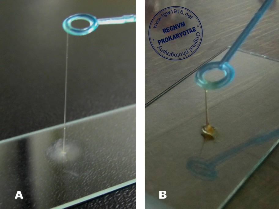
Example 7 - wrong reactives
Indole is a compound that can be produced by
some bacteria as a degradation product of the
amino acid tryptophan. After 24 hours of
incubation, when adding few drops of Kovacs
reagent, it will combine with the indole forming a
pink-red compound.
Reaction is positive if a red layer appears on the
surface of the medium (fig. A). Some bacteria
like Pasteurella can produce a weak reaction
(fig. B).
Fig. C shows what happened when the Kovacs
reagent was replaced by mistake with another
substance (KOH). Red ring was produced in an
indole-negative tube (yet the hue is slightly
modified).
Example 8 - destroyed red cells
Some bacteria have the ability to produce toxins
that lyse erythrocytes from the culture substrate
(blood agar), resulting a greenish or clear halo
surrounding the colonies (partial or complete
hemolysis). The red cells are easily destroyed
by other factors than bacterial activity, like pH
value or salinity of the inoculum.
In this example a false hemolysis is produced by
a low-pH inoculum (C).
Example 9 - serum interference
Horse serum contains a maltase which breaks
down maltose to glucose. Adding horse serum in
medim when testing maltose fermentation may
lead to a false-positive result if the strain
ferments glucose.
Example 10 - acidic gas
The carbon dioxide (CO2) gas dissolved in water
can cause water to become acidic, hence can
change the color of a pH indicator. Fermentation
tests are based on lowering the pH due to acidic
metabolic compounds resulting from the
degradation of sugars by bacteria.
In API tests the pH indicator is turning yellow if
the sugars are fermented. But, a false positive
result may appear if the acidic environment is
caused by a rich carbon dioxide atmosphere. In
this example raffinose (RAF), xylose (XYL) and
methyl-alpha-D-glucopyranoside (MDG) are
negative if the strip is incubated aerobically (A)
and false-positive when incubated in 20% CO2
atmosphere (B).
Example 11 - anaerobiosis issue
Some bacteria require anaerobic conditions for
growth, which can be achieved by adding
paraffin oil to the medium surface which stops
the solubilization of atmospheric oxygen. In the
attached example, the paraffin oil was
mistakenly replaced with glycerin, which look the
same but it is soluble in water and does not
create a film on the surface of the medium.
Therefore, false negative results were obtained
(bottom) compared to tests where paraffin oil
was used (top) in which arginine dihydrolase
(ADH), ribose fermentation (RIB) and trehalose
fermentation (TRE) are positive.
Klebsiella oxytoca strains can produce a dark
brown pigment when growth on media containing
gluconate and ferric citrate. Klebsiella does not
produce hydrogen sulfide and hence does not
produce a dark ring when tested on TSI agar. In
this case the TSI slant is darkened by pigment,
not by hydrogen sulfide production.
Example 2 - pigment on chromogenic media
MacConkey Agar is frequently used as selective
media for the Enterobacteriaceae. The growth of
Gram-positive germs is inhibited by the bile salts
and the crystal violet. Medium also contains
lactose and neutral red as pH indicator.
Lactose-fermenting germs form red colonies,
lactose-negative germs form colorless colonies.
Serratia marcescens does not ferment lactose,
hence it should form colorless colonies. But this
species produce a red pigment resulting red
colonies on MacConkey agar which may be
taken as lactose-positive.
Example 3 - old culture
With very few exceptions, Salmonella is a
microorganism that does not ferment lactose.
Therefore, when grown on MacConkey, it
produces colorless colonies.
In the presented case, following a prolonged
storage in the refrigerator (more than a month),
the environment around the colonies became
acidic and caused the red color change of the
colonies.
Example 4 - blood interference
Catalase is an enzyme present in most of the
organisms and is involved in decomposition of
the hydrogen peroxyde in H2O and O2. If
catalase is present, bubbles will form from the
oxygen that is made in the reaction: 2H2O2 +
catalase => 2H2O + O2 + catalase. Test is
negative if effervescence is not produced.
But not only bacteria can produce catalase;
media containing whole red blood cells will
contain catalase and could therefore give a
false positive result. If using colonies from a
blood agar plate, be very careful not to scrape
up any of the blood agar.
Example 5 - slime
Like the Gram stain reaction, the potassium
hydroxide test is based on differences in the
bacterial wall composition. The cell wall of
Gram-negative bacteria is easily disrupted when
exposed to dilute alkali solutions while the tough
thick peptidoglycan wall of Gram-positives does
not lyse. When the cell walls are disrupted, the
suspension in KOH becomes viscous due to the
release of relatively unfragmented threads of
deoxyribonucleic acid. But, some Gram-positive
species like Bacillus subtilis can produce a
viscous substance / slime resembling the DNA
released from Gram-negatives in KOH solution.
Example 6 - bad neighbours
The results of reactions based on color change
following acidification or alkalinization of the
environment can be falsified by the reagents
added to the nearby tests. In this example of API
20 E kit, 10 minutes after adding the acetic acid
(NIT1 reactive) in the GLU tube, the MAN test
starts to change color to yellow due pH lowering
induced by the acid vapours.
Example 7 - wrong reactives
Indole is a compound that can be produced by
some bacteria as a degradation product of the
amino acid tryptophan. After 24 hours of
incubation, when adding few drops of Kovacs
reagent, it will combine with the indole forming a
pink-red compound.
Reaction is positive if a red layer appears on the
surface of the medium (fig. A). Some bacteria
like Pasteurella can produce a weak reaction
(fig. B).
Fig. C shows what happened when the Kovacs
reagent was replaced by mistake with another
substance (KOH). Red ring was produced in an
indole-negative tube (yet the hue is slightly
modified).
Example 8 - destroyed red cells
Some bacteria have the ability to produce toxins
that lyse erythrocytes from the culture substrate
(blood agar), resulting a greenish or clear halo
surrounding the colonies (partial or complete
hemolysis). The red cells are easily destroyed
by other factors than bacterial activity, like pH
value or salinity of the inoculum.
In this example a false hemolysis is produced by
a low-pH inoculum (C).
Example 9 - serum interference
Horse serum contains a maltase which breaks
down maltose to glucose. Adding horse serum in
medim when testing maltose fermentation may
lead to a false-positive result if the strain
ferments glucose.
Example 10 - acidic gas
The carbon dioxide (CO2) gas dissolved in water
can cause water to become acidic, hence can
change the color of a pH indicator. Fermentation
tests are based on lowering the pH due to acidic
metabolic compounds resulting from the
degradation of sugars by bacteria.
In API tests the pH indicator is turning yellow if
the sugars are fermented. But, a false positive
result may appear if the acidic environment is
caused by a rich carbon dioxide atmosphere. In
this example raffinose (RAF), xylose (XYL) and
methyl-alpha-D-glucopyranoside (MDG) are
negative if the strip is incubated aerobically (A)
and false-positive when incubated in 20% CO2
atmosphere (B).
Example 11 - anaerobiosis issue
Some bacteria require anaerobic conditions for
growth, which can be achieved by adding
paraffin oil to the medium surface which stops
the solubilization of atmospheric oxygen. In the
attached example, the paraffin oil was
mistakenly replaced with glycerin, which look the
same but it is soluble in water and does not
create a film on the surface of the medium.
Therefore, false negative results were obtained
(bottom) compared to tests where paraffin oil
was used (top) in which arginine dihydrolase
(ADH), ribose fermentation (RIB) and trehalose
fermentation (TRE) are positive.
In current microbiological practice, the next step after strain isolation is the identification by
biochemical tests. These tests mostly consist of color reactions based on the synthesis of a
colored metabolic compound, or the pH modification of the culture medium, which in the
presence of a color indicator (bromtymol blue, phenol red) modifies its optical properties
resulting a color change of the medium (eg. alkalinization by lysine decarboxylase or
acidification by sugars fermentation).
Interpretation of these tests is often easy, but in some cases, such as late reading of the
tests, prolonged incubation, the mistaken reagents added, contamination, or the influence
of other neighboring tests, an inexperienced microbiologist can be fooled.
Here are a few examples of false-positive reactions that I have encountered in the lab:
| False-positive H2S production (weak blackening) on TSI medium (left). |
| MacConkey agar: lactose-positive colonies (up), lactose-negative colonies (left) and false lactose-positive, red-pigmented Serratia colonies (right) |
| False lactose-positive colonies on MacConkey agar (Salmonella serovar Typhimurium) |
| A - KOH test revealing the Gram-negative wall structure of Escherichia coli, B - slime produced by Bacillus subtilis - false KOH result. |
| Negative MAN (mannitol fermenting) test starting to become false-positive after adding NIT1 reactive in GLU test |


|


| False-negative results in API Strep tests due to replacing paraffin oil with glycerin (down). Correct results in the above strip. |