| S. epidermidis haemolytic strain |
| Staphylococcus epidermidis |
| S. epidermidis cells, Gram-stained |
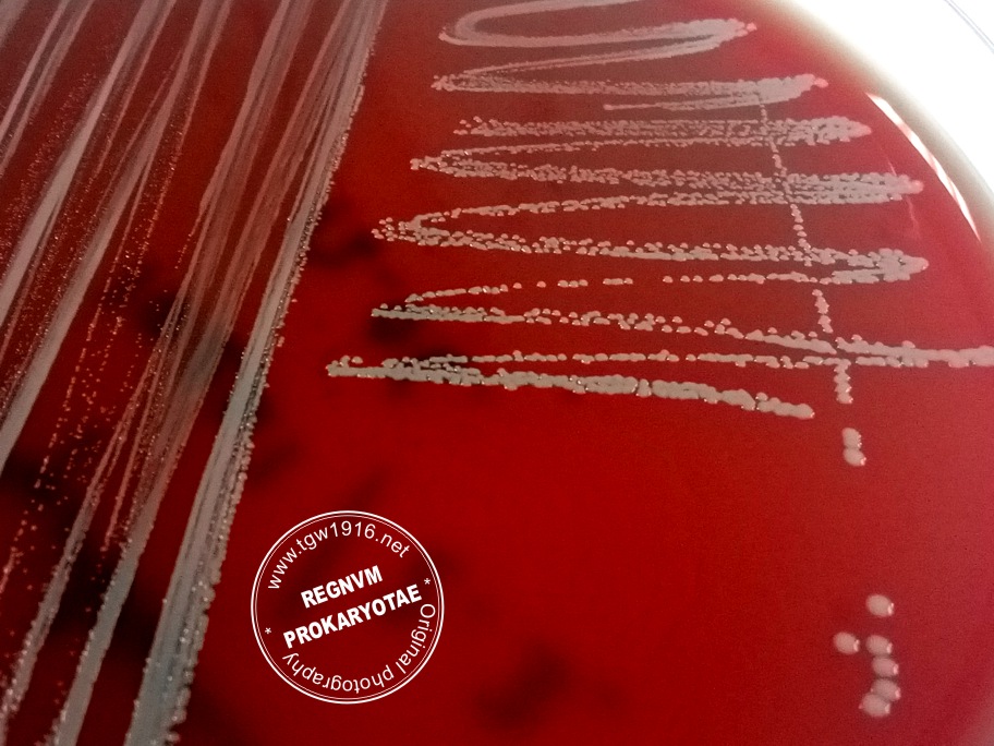
| S. epidermidis colonies on Sheep Blood Agar |
Taxonomy
Morphology
Cultural characteristics
Biochemical characters
Ecology
Pathogenicity
References
Phylum Firmicutes, Class Bacilli, Order Bacillales, Family Staphylococcaceae, Genus Staphylococcus, Staphylococcus epidermidis
Evans 1916.
Old synonyms: Albococcus epidermidis Winslow and Winslow 1908; Micrococcus epidermidis Hucker 1924.
Evans 1916.
Old synonyms: Albococcus epidermidis Winslow and Winslow 1908; Micrococcus epidermidis Hucker 1924.
Gram-positive cocci, 0.5-1.5 μm, nonmotile, nonsporing. Occur in pairs and tetrads.
Colonies are small, white or yellow (usually nonpigmented - gray or grayish white),
approximately 2.5-4 mm in diameter, usually nonhemolytic. Growth in broth is first
turbid, later becoming clear with a fine or slightly mucoid deposit. Facultatively
anaerobic, optimum temperature 26-37 ºC (growth range 15-45 ºC). Grows on media:
Trypticase Soy Agar ± 5% sheep blood, Chapman (selective medium with 75 g/l NaCl
and mannitol), Mueller-Hinton agar.
Isolated from human and animal skin and mucous membranes. Isolated from bovine
milk (admin note). Found in fermenting cigar tobacco leafs. Susceptible to Novobiocin.
milk (admin note). Found in fermenting cigar tobacco leafs. Susceptible to Novobiocin.
Usually nonpathogenic to humans, may be an important cause of infection with
compromised immunity, also a nosocomial pathogen associated with infections of
implanted medical devices. Produces a slime resulting in biofilm formation on the
surface of a prosthetic device (catheter). Bacteremia, endophtalmitis and endocarditis.
Bacteria is a pig pathogen causing contagious impetigo in swine.
Subclinical mastitis in goats. Subclinical mastitis in cows (admin note).
Produces the antibiotic ‘Epidermin’.
- Holt J.G., Krieg N.R., Sneath P.H.A., Staley J.T., Wiliams S.T., 1994. Bergey's
Manual of Determinative Bacteriology, Ninth Edition, Williams & Wilkins,
Baltimore. Group 17, Gram-Positive Cocci, 527-558. - Evans A.C.: The bacteria of milk freshly drawn from normal udders. Journal of
Infectious Diseases, 1916, 18, 437-476. - Jerome J. Perry: Isolation of Staphylococcus epidermidis from Tobacco. Applied
Microbiology, Apr. 1969, p. 647. - P. Moroni, G. Pisoni, M. Antonini, G. Ruffo, S. Carli, G. Varisco and P. Boettcher:
Subclinical Mastitis and Antimicrobial Susceptibility of Staphylococcus caprae and
Staphylococcus epidermidis Isolated from Two Italian Goat Herds. J. Dairy Sci. 88:
1694-1704. - Karl-Heinz Schleifer and Julia A. Bell, 2009. Family VIII. Staphylococcaceae fam.
nov.. In: (Eds.) P.D. Vos, G. Garrity, D. Jones, N.R. Krieg, W. Ludwig, F.A. Rainey, K.
-H. Schleifer, W.B. Whitman. Bergey’s Manual of Systematic Bacteriology, Volume
3: The Firmicutes, Springer, 392-426. - Okee MS, Joloba ML, Okello M, Najjuka FC, Katabazi FA, Bwanga F, Nanteza A,
Kateete DP. Prevalence of virulence determinants in Staphylococcus epidermidis
from ICU patients in Kampala, Uganda. J Infect Dev Ctries. 2012 Mar 12;6(3):242-
50. doi: 10.3855/jidc.2007. PMID: 22421605.
Positive results for nitrate reduction, alkaline phosphatase, arginine dihydrolase,
urease, acetoin production, catalase, acid production from mannose and sucrose.
Negative results for starch hydrolysis, oxidase, growth on (NH4)2SO4, coagulase-rabbit
plasma, clumping factor, ornithine decarboxylase, deoxyribonuclease agar,
heat-stable nuclease, beta-glucuronidase, beta-galactosidase (most strains), acid
production from erythritol, erythrose, gentiobiose, inositol, lyxose, sorbose, tagatose,
trehalose, mannitol, xylitol, raffinose, arabinose, cellobiose, fucose, and salicin.
Variable results for hyaluronidase, beta-glucosidase, acid production from lactose,
galactose, melezitose, turanose and ribose.

(c) Costin Stoica

| Antibiogram |
| Encyclopedia |
| Culture media |
| Biochemical tests |
| Stainings |
| Images |
| Movies |
| Articles |
| Identification |
| Software |
| R E G N U M PROKARYOTAE |

| Back |