
| Listeria monocytogenes |
Facultative anaerobic. Organic growth factors are required. Grow at/on: pH 5.5-9.6 (pH 7.2-7.6 optimum). Temperature range 0-45 ºC.
Grow on 40% bile agar. Weak growth on MacConkey agar. Variable growth in 20% NaCl or in peptone water plus 10% NaCl.
Grow on 40% bile agar. Weak growth on MacConkey agar. Variable growth in 20% NaCl or in peptone water plus 10% NaCl.
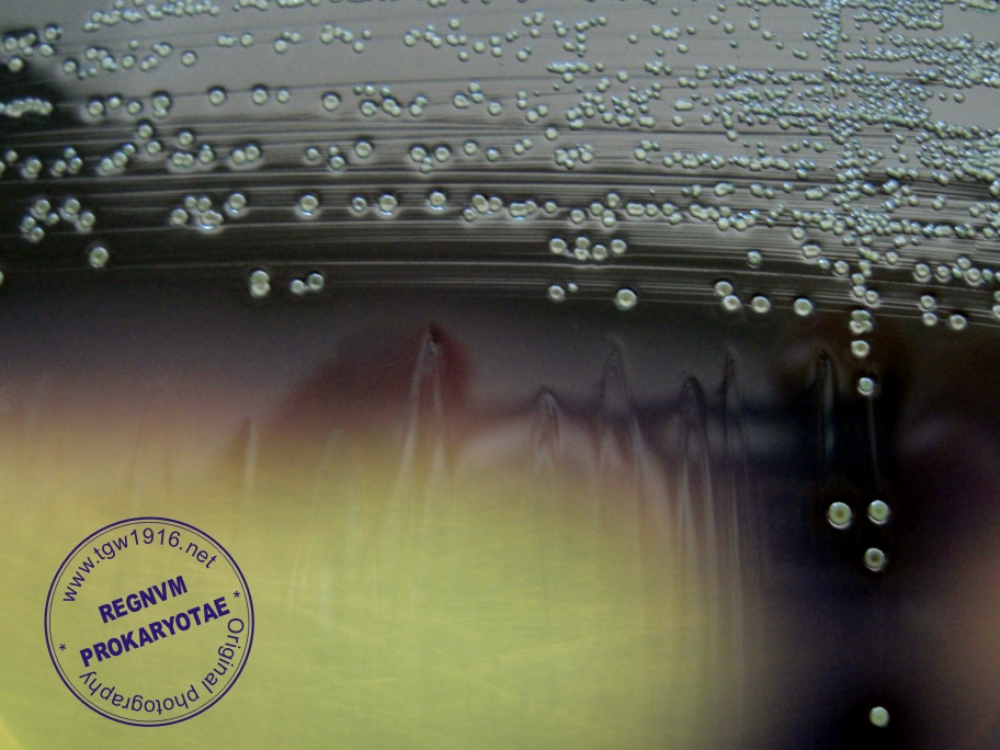
| Listeria monocitogenes colonies on Oxford Agar |
Taxonomy
Morphology
Cultural characteristics
Biochemical characters
Ecology
Pathogenicity
References
Phylum Bacillota (Firmicutes), Class Bacilli, Order Bacillales, Family Listeriaceae, Genus Listeria, Listeria monocytogenes (Murray,
Webb and Swann 1926) Pirie 1940.
L. monocytogenes has 13 serovars (1/2a, 1/2b, 1/2c, 3a, 3b, 3c, 4a, 4ab, 4b, 4c, 4d, 4e, 7), with 13 somatic (O factor) antigens (I-XIII)
and 4 flagella (H factor) antigens.
Historical synonyms: Bacterium monocytogenes Murray, Webb and Swann 1926, Listerella hepatolytica Pirie 1927, Bacterium
monocytogenes hominis Nyfeldt 1932, Corynebacterium parvulum Schultz, Terry, Brice and Gebhardt 1934, Erysipelothrix
monocytogenes (Murray et al. 1926) Wilson and Miles 1946, Corynebacterium infantisepticum Potel 1950.
Gram-positive (sometimes Gram-negative, especially in older cultures) short rods or
coccobacilli, 0.4-0.5/1-3 µm, with rounded ends. Coccoid forms are sometimes seen
in smears from infected tissue or from liquid cultures. Usually occur singly or in short
chains, V- or Y-shaped or parallel pairs. In older or rough cultures, and in media
containing high levels of NaCl, filaments up to 96 μm in length may develop.
Nonspore-forming, nonencapsulated.
Motile by 2-6 peritrichous flagella when cultured below 30 ºC but over 20 ºC (L.m. type
strain is nonmotile!) . Expression of the structural gene for the flagellin protein (flaA)
has been shown to be temperature regulated and repressed at 37 ºC. High-level
expression is seen at 25 ºC, corresponding to the temperature at which tumbling
motility is observed. Even in the absence of active motility, strong induction of flagella
biosynthesis occurs below 20 ºC and flagellae have a role in attachment to solid
surfaces. Motility can also be demonstrated in semisolid agar in “U” tubes. Maximal
turbidity in liquid and semisolid agar is present 2-3 mm below the surface in an
umbrella-like pattern.
Grow well on blood agar base, nutrient, tryptose, and tryptose soy or brain heart
infusion agars; growth is enhanced by the addition of 0.2–1% (w/v) glucose, blood or
serum. For specimens such as feces, vaginal secretions, food and environmental
samples, special selective media are necessary: these include acriflavin, lithium
chloride, colistin, ceftazidime, cefotetan, fosfomycin, moxolactam, nalidixic acid,
cycloheximide, and polymyxin.
On solid media containing blood, overnight, the colonies are translucent, slightly
raised, non-pigmented, 1-2 mm in diameter with a ‘ground-glass’ crystalline
appearance in the center, have a entire margin and a watery consistency and easy
emulsifiable. The rough forms have larger, flatter colonies, with an irregular indented
edge and are umbonate with a central crater, friable, and difficult to emulsify. All
cultures have a characteristic sweet, buttermilk-like smell.
Growth in broth is poor, but is improved by the addition of 0.5-1.0 % glucose; in this
medium the smooth forms give rise to a fairly dense suspension, and after some
days the organisms settle to the bottom in floculles.
Beta-hemolytic on agars containing sheep, cow, horse, rabbit or human blood and
CAMP-test-positive with Staphylococcus aureus and negative with Rhodococcus equi
(L. m. type strain is nonhemolytic and CAMP-test-negative!).
Widely distributed in nature, found in soil, fresh water, wastewater, mud, sewage,
vegetation, fresh and frozen poultry, slaughterhouse wastes and in the feces of
animals and man; there is an asymptomatic carriage in the intestinal tract. It can also
colonize various inert surfaces and can form biofilms on food-processing surfaces.
Multiply rapidly in milk. Grow in the presence of 10 µg/ml trypaflavine, 0.025% (w/v)
thallous acetate, 3.75% (w/v) potassium thiocyanate or 0.04% (w/v) potassium
tellurite. No growth in the presence of 0.02% (w/v) sodium azide.
Plasmids encoding resistance to tetracycline alone and multiresistance to
chloramphenicol, erythromycin, streptomycin and tetracycline have been detected,
although these are rare.
Is usually sensitive to amikacin, amoxycillin, ampicillin, azlocillin, ciprofloxacin,
chloramphenicol, clindamycin coumermycin, doxycycline, enoxacin, erythromycin,
gentamicin, imipen, netilmicin, penicillin, rifampin, trimethoprim, and vancomycin.
Resistant to the cephalosporins, phosphomycin, and polymyxin.
Lytic properties of sets of phages have been used for subtyping L. monocytogenes.
Bacteria are killed at 59-63 ºC in 10 minutes and pasteurization is a good technique
to eliminate the bacteria from dairy products.
Listeria monocytogenes is a facultatively intracellular pathogen for a wide variety of animals (mammals, birds, fish and invertebrates)
and man, causing septic lesions in various organs (uterus at pregnancy, central nervous system, etc: gastroenteritis, meningitis,
neuro-encephalitis, endocarditis, chorioamnionitis, septicaemia, abortions and neonatal infections); a disease named listeriosis.
Most cases are food- or feed-borne. The majority of strain causing disease (> 90%) in humans and animals belong to serovars 1/2a
(especially from animals), 1/2b, and 4b (especially from humans)
For considerations for public-health purposes, all strains of L. monocytogenes should be regarded as potentially pathogenic (non-
pathogenic variants have been described and there may be differences in virulence between strains).
A wide range of animals are susceptible to experimental infection, but rabbits, mice and, to a lesser extent, rats are most frequently
used since these animals die following inoculation of live bacteria by the intravenous or intraperitoneal route. The production of
experimental keratoconjunctivitis (Anton’s eye test) in guinea pigs or rabbits by instilling a live bacterial suspension into the
conjunctiva, historically has been used to demonstrate the virulence of L. monocytogenes. A purulent conjunctivitis and occasionally a
keratitis develops within 24-48 h; usually heals spontaneously. Pathogenic for mice.
Repression of flagellae at 37 ºC is necessary for full virulence. Cell surface and secreted proteins occur, some of which are involved
with the virulence.
The production of many of the surface-associated virulence genes is temperature-dependent and these are expressed above 30 ºC.
Show properties of invasion and spreading in a range of mammalian cells growing in vitro. Enters both phagocytic and non-
phagocytic cells. A listerial surface protein, internalin A (which is reminiscent of the M protein of the group A streptococci), has been
shown to be involved with the initial stages of invasion of all cell types. A second cell surface protein (internalin B) is required for entry
into hepatocyte-like but not into enterocyte-like cells. The cell-wall teichoic acid is also required for adhesion of Listeria to mammalian
cells. The listerial cell-surface protein, p60 (with murein hydrolase activity) may also be involved in the invasion of fibroblasts and a
second surface-exposed autolysin (Ami) has been shown to be involved in adhesion to eukaryotic cells. Intracellular motility is
promoted by the ActA protein; this protein is involved in intercellular spreading too.
Listeriolysin O, a thiol-activated hemolysin, mediate the dissolution of the phagocytic intracellular vacuole membrane, in combination
with the action of a phospholipase C.
Virulence for Listeria monocytogenes is a multifactorial property and at least nine genes and their products are required for infection,
invasion, survival, mobility and cell-to-cell spread, although additional genes (especially those for cell-surface components) are also
likely to be involved with the infectious process.
- Hammes W.P. and Hertel C., 2009. Genus I. Listeria Pirie 1940. In: (Eds.) P.D. Vos, G. Garrity, D. Jones, N.R. Krieg, W. Ludwig, F.A.
Rainey, K.-H. Schleifer, W.B. Whitman. Bergey’s Manual of Systematic Bacteriology, Volume 3: The Firmicutes, Springer, 244-257. - Raducanescu H. si V. B. Popii, 1986. Genul Listeria. In: Bacteriologie veterinara, Ed. Ceres, Bucuresti, 140-144.
- McLauchlin J., 2005. Listeria. In: Topley & Wilson’s Microbiology & Microbial Infections, 10th Edition, Edited by Borriello S.P., Murray
P.R. and Funke G., 953-969. - Khelef N., Lecuit M., Buchrieser C., Cabanes D., Dussurget O. and Cossart P., 2006. Listeria monocytogenes and the genus
Listeria. In: Dworkin M., Falkow S., Rosenberg E., Schleifer K.H., Stackebrandt (Editors), The Prokaryotes, A Handbook on the
Biology of Bacteria, Third Edition, Volume 4, Bacteria: Firmicutes, Cyanobacteria, Springer, Chapter 1.2.11, 404-476. - Caplan D. M., 2009. Listeria. In: Buiuc D., Negut M. (Editori), Tratat de Microbiologie Clinica, Editia a IIIa, Editura Medicala, Bucuresti,
687-692. - Seeliger H.P.R. and Welshimer H.J., 1974. Genus Listeria Pirie 1940. In: Buchanan R.E. and Gibbons N.E. (Editors), Bergey’s
Manual of Determinative Bacteriology, Eight Edition, The Williams & Wilkins Company, Baltimore, 593-596. - Den Bakker, H. C., Warchocki, S., Wright, E. M., Allred, A. F., Ahlstrom, C., Manuel, C. S., Stasiewicz, M. J., Burrell, A., Roof, S., Strawn,
L. K., Fortes, E., Nightingale, K. K., Kephart, D. and Wiedmann, M. 2014. Listeria floridensis sp. nov., Listeria aquatica sp. nov.,
Listeria cornellensis sp. nov., Listeria riparia sp. nov. and Listeria grandensis sp. nov., from agricultural and natural environments.
Int. J. Syst. Evol. Microbiol., 64, 1882-1889. - Jocelyne Rocourt, Benedicte Catimel. Caracterisation biochimique des especes du genre Listeria, Zentralblatt für Bakteriologie,
Mikrobiologie und Hygiene. Series A: Medical Microbiology, Infectious Diseases, Virology, Parasitology, Volume 260, Issue 2, 1985,
Pages 221-231, ISSN 0176-6724, https://doi.org/10.1016/S0176-6724(85)80118-8.
Positive results for acid phosphatase, catalase (few strain negative), chymotrypsin, esculin hydrolysis, beta-glucosidase, hippurate
hydrolysis, methyl-red, Voges-Proskauer, lecithinase, phosphoamidase, tween 80 esterase, acid production from: esculin, D-glucose,
glycerol, lactose, maltose, methyl alpha-D-glucoside, methyl alpha-D-mannoside, rhamnose, trehalose, turanose, salicin and xylitol.
Negative results for casein hydrolysis, cellulose hydrolysis, citrate utilization, gelatin hydrolysis, H2S production, indole production,
nitrates reduction to nitrites, oxidase, urea hydrolysis, arylesterase activity, cystine arylamidase, amino acid peptidase: D-alanine,
lysine, ornithine, glutamic acid and arginine; acid production from: L-arabinose, gluconate, glucose 1-phosphate, glycogen, D-mannitol,
melibiose, ribose, soluble starch and D-xylose.
Variable results for alpha-glucosidase, N-acetyl-beta-glucosamidase, leucine esterase, starch hydrolysis, acid production from: dextrin,
galactose, 5-ketogluconate, D-lyxose, melezitose, sorbitol, sucrose and tagatose.
(c) Costin Stoica

| Antibiogram |
| Encyclopedia |
| Culture media |
| Biochemical tests |
| Stainings |
| Images |
| Movies |
| Articles |
| Identification |
| Software |
| R E G N U M PROKARYOTAE |


| Back |